Tartaczna 2
Discover the quality of healthcare in the style of Tartaczna2!


 back
back
An expert endometriosis ultrasound is a detailed transvaginal examination used to detect and map changes associated with endometriosis, particularly endometriomas, pelvic adhesions, adenomyosis and deep endometriosis.
At Tartaczna 2 Medical Centre, the expert endometriosis ultrasound in Gdańsk is performed exclusively by Dr Dominika Kozikowska. The examination includes a systematic assessment of the pelvis, and any identified lesions may be mapped and described using the #Enzian classification.
This is a separate, more comprehensive service than a standard gynaecological ultrasound. When booking, please select “endometriosis ultrasound” rather than a standard gynaecological consultation or routine transvaginal ultrasound.
A standard transvaginal ultrasound primarily assesses the uterus, endometrium and ovaries. It may detect fibroids, polyps, ovarian cysts and other abnormalities of the reproductive organs.
An expert endometriosis ultrasound has a considerably broader scope. In addition to assessing the uterus and ovaries, it includes a systematic search for lesions in the anterior and posterior pelvic compartments, an assessment of organ mobility and adhesions, and an examination of structures that may be affected by deep endometriosis.
The examination also includes dynamic ultrasound assessment. The doctor evaluates the movement of the uterus, ovaries, bowel and other pelvic structures in relation to one another. Restricted mobility may indicate adhesions or deep endometriotic lesions.
The examination may be recommended if you experience symptoms or have previous findings that may suggest endometriosis, including:
The severity of pain does not always reflect the extent of endometriosis. Small lesions may cause severe symptoms, while more extensive disease may sometimes cause relatively few symptoms.
The scope of the examination is adapted to the patient’s symptoms and clinical situation. The assessment may include:
An expert ultrasound may help determine the location, size and extent of lesions and their relationship with adjacent organs. This information is important when planning conservative management, further assessment with MRI or possible surgical treatment.
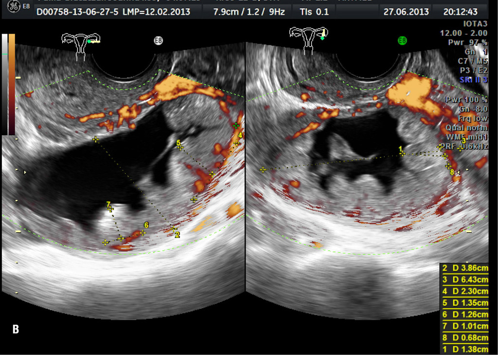
The systematic ultrasound assessment of patients with suspected endometriosis is based on principles developed by the International Deep Endometriosis Analysis group, known as IDEA. It includes examination of the uterus and adnexa, a search for indirect signs of the disease, assessment of organ mobility and evaluation of the anterior and posterior pelvic compartments.
The results may be described using the #Enzian classification. This system provides a structured description of the location and extent of endometriosis, including ovarian involvement, adhesions, deep endometriosis and lesions involving the bowel, urinary bladder or ureters.
#Enzian is a classification used to map the disease rather than a separate type of ultrasound examination. It facilitates communication between the doctor performing the ultrasound, the treating gynaecologist, radiologist and surgical team.
Before the examination, Dr Dominika Kozikowska takes a medical history covering the patient’s symptoms, their relationship with the menstrual cycle, previous treatment, previous operations and plans for pregnancy.
The examination is primarily performed using a transvaginal ultrasound probe. When necessary, the assessment may be supplemented with an abdominal ultrasound.
During the examination, the doctor assesses not only the appearance of individual organs but also their mobility in relation to one another. Gentle probe movements and light pressure may be used in selected areas of the pelvis. This helps assess tenderness, adhesions and the ultrasound sliding sign.
The examination usually takes approximately 30 minutes, although its duration depends on the patient’s anatomy, the severity of symptoms and the extent of any identified lesions.
After the examination, the patient receives a report containing the most important findings. The doctor discusses any abnormalities and explains the possible next diagnostic or treatment steps.
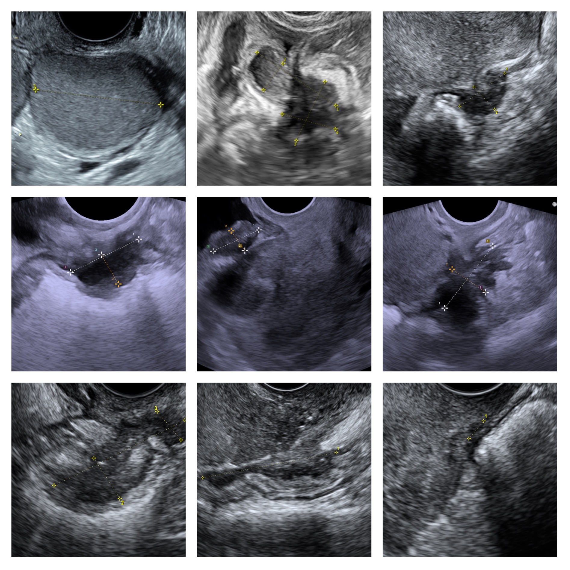
Examples of areas assessed during an endometriosis ultrasound examination.
The examination does not require surgery, but it may cause temporary discomfort in patients with severe pelvic pain or deep endometriosis. Areas affected by the disease may be particularly sensitive during assessment.
The examination is performed with consideration for the patient’s comfort. You can report pain, ask the doctor to reduce the pressure or request that the examination be stopped at any time.
Preparation may depend on the planned scope of the examination. Detailed instructions are provided when the appointment is booked.
Please bring any relevant medical records, including:
Do not take laxatives or perform an enema unless this preparation has been specifically recommended by our reception team or doctor.
The examination can usually be performed on any day of the menstrual cycle. If you are experiencing heavy bleeding, think you may be pregnant or have any questions about preparation, please contact our reception team before the appointment.
A normal expert ultrasound result does not exclude every form of endometriosis. The examination can identify endometriomas and many forms of deep endometriosis with good accuracy, but small superficial peritoneal lesions may remain invisible on imaging.
The ultrasound result should always be interpreted together with the patient’s symptoms, medical history and gynaecological examination. If symptoms persist despite a normal ultrasound, the doctor may recommend monitoring, medical treatment, an MRI scan or another form of diagnostic assessment.
Detecting endometriosis on ultrasound does not automatically mean that surgery is required. The decision depends on the nature and severity of symptoms, the location of the lesions, the patient’s age, plans for pregnancy and the effectiveness of previous treatment.
Medical treatment and monitoring may be appropriate for some patients. Surgery may be considered in cases of severe symptoms, unsuccessful conservative treatment, selected fertility problems or lesions affecting the function of the bowel or urinary system.
Depending on the ultrasound findings and the patient’s clinical situation, the doctor may recommend additional investigations:
CA-125 and CA 19-9 are not sufficiently specific to confirm or exclude endometriosis on their own. They do not replace a medical consultation or appropriately performed diagnostic imaging.
Expert endometriosis ultrasound scans at Tartaczna 2 Medical Centre are performed exclusively by Dr Dominika Kozikowska. The examination includes a detailed assessment of the pelvis and mapping of identified lesions, which may be described using the #Enzian classification.
Due to the specialist nature of the examination and high demand, we recommend booking in advance. Our reception team can provide information about the earliest available appointment and the possibility of joining the waiting list.
Book the examination through our contact form or telephone reception service, call +48 58 719 10 25 or book through ZnanyLekarz: Dr Dominika Kozikowska.
For more information about symptoms and treatment, visit: endometriosis in Gdańsk – symptoms, diagnosis and treatment.