Tartaczna 2


 back
back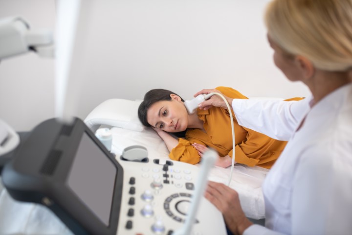
Lymph node ultrasound
Assessment of lymph nodes in the neck, armpits and groin
Lymph node ultrasound helps assess palpable lumps and enlarged lymph nodes and determine whether further investigations are needed. At Tartaczna 2 Medical Centre in Gdańsk, examinations are performed by radiologist Dr Ewa Kołyszko. Ultrasound can assess accessible superficial lymph nodes, including those in the neck, armpits and groin.
Why do lymph nodes become enlarged?
Lymph nodes are part of the immune system. They may swell during an infection, such as a throat, dental or skin infection, or after a vaccination. Other causes include inflammatory and autoimmune conditions, lymphoma and cancer that has spread to lymph nodes. Enlargement alone does not establish the cause.
When should enlarged lymph nodes be assessed?
- A node is growing, remains enlarged or has no clear link to an infection.
- A lump feels hard, does not move freely or is located above the collarbone.
- Lymph nodes are enlarged in several areas of the body.
- There is unexplained fever, night sweats or unintentional weight loss.
- Follow-up has been recommended after a previous examination or treatment.
The doctor first reviews your symptoms and medical history. Not every swollen node during a short-lived infection needs an immediate ultrasound. Difficulty breathing or significant difficulty swallowing requires urgent medical attention.

What does the doctor assess on ultrasound?
The radiologist examines the size, shape, internal structure, cortex, hilum and blood flow of the lymph nodes. Their location and overall appearance matter. There is no single size threshold that can reliably classify every node as normal or cancerous.
The term “reactive lymph nodes” describes an appearance suggesting a response to inflammation or other immune stimulation. Suspicious ultrasound features warrant further assessment; they do not by themselves confirm cancer. The findings must be considered alongside the clinical examination and other investigations.
How to prepare, and what happens during the scan
Ultrasound of superficial lymph nodes usually requires no fasting or changes to your regular medicines. Bring previous results and tell the doctor about recent infections, vaccinations and treatment. When booking, specify which area needs to be examined.
The doctor applies gel and moves a probe over the skin. We use a GE LOGIQ E10 ultrasound system. The scan involves no needle insertion or X-rays. Pressure over a tender area may cause discomfort.
What happens after a lymph node ultrasound?
Depending on the findings, the doctor may recommend observation, blood tests, another imaging examination or a biopsy. Ultrasound does not assess every lymph node in the body and may not show deeply located nodes adequately.
Fine needle aspiration provides cells for cytological examination. If lymphoma is suspected or the result is inconclusive, a core needle biopsy or surgical removal of a node for histopathological examination may be needed. The doctor managing your assessment selects the appropriate sampling method.
Lymph node ultrasound in Gdańsk – appointments
Examinations are performed by Dr Ewa Kołyszko.
Book using the appointment calendar below or contact our reception team: +48 58 719 10 25, kontakt@tartaczna2.pl.