
Tartaczna 2

-
Home

-
OFFER
-
Obstetrics and Gynecology Gdańsk
- Prenatal testing - dual test (USG with risk assessment according to FMF)
 back
back
Prenatal testing - dual test (USG with risk assessment according to FMF)
First-Trimester Prenatal Screening - what Does the Examination Involve?
At Tartaczna 2 Medical Centre, we perform prenatal screening in Gdańsk using the advanced GE Voluson Signature 18 ultrasound system.
This latest-generation ultrasound system provides high-resolution imaging, advanced 3D and 4D fetal visualisation, and the option to save images and video recordings from the examination.
Tartaczna 2 Medical Centre also provides comprehensive antenatal care. Our multidisciplinary team, led by Dr Marek Korożan, a gynaecologist and obstetrician, includes experienced doctors, midwives, an endocrinologist, a diabetologist and an on-site laboratory. Together, we support the healthy progress of pregnancy and look after the well-being of both mother and baby.
Prenatal screening – why is it important?
Modern prenatal care includes screening tests and ultrasound examinations that can assess the chance of certain chromosomal conditions and detect some structural fetal abnormalities at an early stage of pregnancy.
Combined first-trimester screening is used primarily to assess the chance of the most common trisomies: Down's syndrome (trisomy 21), Edwards' syndrome (trisomy 18) and Patau's syndrome (trisomy 13). It includes a specialist ultrasound scan, measurement of biochemical markers in the mother's blood and analysis of relevant maternal characteristics.
Ultrasound and blood sampling are non-invasive procedures. They are not associated with the risk of miscarriage that applies to invasive diagnostic procedures. It is important to remember, however, that screening estimates the chance of a particular condition and does not provide a diagnosis.
First-trimester prenatal screening – the combined test and double test
Combined first-trimester screening takes into account the results of a specialist ultrasound examination, relevant maternal characteristics and the levels of pregnancy-associated plasma protein A (PAPP-A) and free beta-human chorionic gonadotropin (free β-hCG) in the mother's blood.
The term “double test” refers specifically to the biochemical component, involving measurement of two markers: PAPP-A and free β-hCG. The results of the blood tests must be combined with the ultrasound findings and maternal characteristics to calculate the individual chance of fetal trisomy.
A higher-chance result does not mean that a chromosomal condition has been diagnosed in the baby. It indicates that the result should be discussed with a doctor and that further screening or diagnostic testing may be considered.

When should first-trimester prenatal screening be performed?
An FMF-standard first-trimester ultrasound scan is performed between 11 weeks and 13 weeks plus 6 days of pregnancy, when the fetal crown–rump length (CRL) measures between 45 and 84 mm.
During this period, standardised ultrasound measurements can be obtained, the early fetal anatomy can be assessed and the individual chance of the most common trisomies can be calculated. The timing of the blood sample for the double test is determined by the doctor or laboratory. The sample may be collected before the ultrasound scan or on the day of the examination.
PAPP-A test – maternal blood test
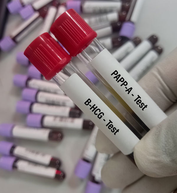
The biochemical component of combined first-trimester screening measures the levels of pregnancy-associated plasma protein A (PAPP-A) and free beta-human chorionic gonadotropin (free β-hCG) in the mother's blood.
The levels of both markers are adjusted for gestational age and selected maternal characteristics. They should not be interpreted separately. Combining the biochemical results with the ultrasound measurements and maternal characteristics provides a more reliable assessment of the chance of fetal trisomy.
First-trimester prenatal ultrasound – what does the doctor assess?
During a first-trimester prenatal scan, the doctor assesses fetal development, confirms the gestational age and examines the anatomical structures and ultrasound markers that can be visualised at this stage. The exact scope of the examination depends on factors including gestational age, fetal position and imaging conditions.
Nuchal translucency (NT)
Nuchal translucency, abbreviated to NT, is the ultrasound appearance of a fluid-filled space beneath the skin at the back of the baby's neck.
NT measurement is one of the most important components of first-trimester screening for chromosomal conditions. Increased nuchal translucency may be associated with a higher chance of trisomy, congenital heart defects and certain other syndromes or developmental abnormalities. It does not, however, provide a diagnosis on its own.
Crown–rump length (CRL)
The fetal crown–rump length, or CRL, is measured to confirm the gestational age and estimated due date. An accurate CRL measurement is also essential for the correct interpretation of nuchal translucency and double-test results.
Early assessment of fetal anatomy
During the ultrasound examination, the doctor assesses the fetal anatomical structures that can be visualised at this stage. These may include the skull and brain, facial profile, spine, chest, heart, abdominal wall, stomach, urinary bladder and limbs.
The nasal bone may also be assessed as an additional ultrasound marker. An absent or hypoplastic nasal bone may be associated with a higher chance of certain chromosomal conditions, but this finding alone does not provide a diagnosis.
Ductus venosus flow and fetal heart assessment
When clinically appropriate, the examination may be extended to include Doppler assessment of blood flow through the ductus venosus and across the tricuspid valve. The doctor also assesses the fetal heart rate and the cardiac structures that can be visualised at this stage.
Abnormal ductus venosus flow or tricuspid regurgitation may be associated with a higher chance of trisomy or congenital heart defects. These findings must be interpreted together with the other components of the examination and cannot independently confirm or exclude a heart defect.

Understanding prenatal screening results
Specialist software is used to calculate the combined screening result. The analysis takes into account maternal age and other relevant maternal characteristics, PAPP-A and free β-hCG levels, CRL and NT measurements, and any additional ultrasound markers that have been assessed.
The result is expressed as an individual chance of a particular trisomy, for example 1 in 1,000 or 1 in 50. Depending on the calculation method and the applicable thresholds, the result may be classified as lower, intermediate or higher chance.
A lower-chance result is reassuring and means that the assessed condition is less likely. It does not, however, guarantee a normal fetal karyotype or exclude every genetic condition or developmental abnormality.
A higher-chance result does not mean that the baby has been diagnosed with a condition. It should be discussed with a doctor, who will explain its significance and the available options for further testing.
FMF standards – what do they mean?
The FMF, or The Fetal Medicine Foundation, is an international organisation involved in scientific research, education, certification and the development of methods used in fetal medicine.
FMF protocols define, among other things, how ultrasound measurements should be obtained, the conditions required for assessing first-trimester markers and the methods used to calculate the chance of fetal trisomy. The use of standardised methodology improves the reproducibility of measurements and reliability of the risk calculation.
Prenatal screening should be performed by a suitably trained doctor using appropriate ultrasound equipment. Advanced ultrasound technology alone cannot replace the doctor's experience or the correct interpretation of all the examination results.
How is prenatal ultrasound performed – transabdominally or transvaginally?
A first-trimester ultrasound examination is usually performed transabdominally. Ultrasound gel is applied to the patient's abdomen, and the doctor obtains the required images using an abdominal transducer.
If the fetal position or imaging conditions do not allow all the required structures to be assessed accurately, the doctor may propose an additional transvaginal scan. The choice of method depends on the clinical situation, the quality of the images obtained and the patient's consent.

Higher-chance prenatal screening result – what happens next?
If combined screening indicates a higher chance of a chromosomal condition, the doctor will discuss the significance of the result and the available options with the patient. Depending on the individual circumstances, these may include a specialist consultation, a more detailed ultrasound scan, non-invasive prenatal testing (NIPT) or a diagnostic test.
NIPT, including the NIFTY and Harmony tests, analyses cell-free DNA of placental origin present in the mother's blood. It is a more accurate screening test for selected trisomies, but it is not a diagnostic test. A higher-chance NIPT result should be confirmed by invasive diagnostic testing.
Invasive diagnostic tests include chorionic villus sampling (CVS) and amniocentesis. They allow laboratory analysis of material obtained from the placenta or amniotic fluid. The choice of test depends on factors including gestational age, ultrasound findings and the type of suspected condition. Its potential benefits and risks should be discussed individually with a doctor.
Prenatal screening and first-trimester ultrasound in Gdańsk
At Tartaczna 2 Medical Centre in Gdańsk, we provide comprehensive prenatal assessment, including combined first-trimester screening, PAPP-A testing and specialist prenatal ultrasound examinations.
Our gynaecologists and obstetricians use advanced ultrasound equipment and standardised prenatal screening protocols. Each result is discussed individually with the patient, together with an explanation of its significance and recommendations regarding any further tests that may be appropriate.
We invite you to book an appointment with one of our gynaecologists and obstetricians in Gdańsk.



Pregnancy management and prenatal care

Midwife consultation and lactation outpatient clinic
