
Tartaczna 2


 back
back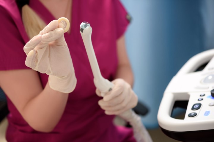
Gynaecological Ultrasound Gdańsk – Transvaginal Scan
Ultrasound assessment of the uterus, endometrium and ovaries
Gynaecological ultrasound in Gdańsk, including transvaginal ultrasound, is one of the main imaging examinations used in gynaecological diagnosis. It allows the doctor to assess the uterus, endometrium, ovaries and other pelvic structures that can be visualised during the scan.
At Tartaczna 2 Medical Centre, the examination is performed by a gynaecologist. Positioning the probe close to the pelvic organs usually provides a more detailed view than a transabdominal scan. A transvaginal ultrasound is generally performed with an empty bladder.
What is a transvaginal ultrasound?
A transvaginal ultrasound is an internal pelvic ultrasound examination. It may also be called a vaginal ultrasound, TVUS or TVS. The probe is gently inserted into the vagina, where it produces images of the pelvic organs using sound waves.
The term “intrauterine ultrasound” is not an accurate description of a standard transvaginal scan. The probe remains in the vagina and is not inserted into the uterine cavity.
When is a gynaecological ultrasound recommended?
A gynaecological ultrasound may form part of a consultation or be arranged to investigate particular symptoms or monitor a previously identified condition. Common reasons for the examination include:
- lower abdominal pain or chronic pelvic pain,
- heavy, painful or irregular periods,
- bleeding or spotting between periods,
- bleeding after the menopause,
- suspected fibroids, endometrial polyps or adenomyosis,
- a suspected ovarian cyst or another adnexal mass,
- monitoring previously identified changes in the uterus or ovaries,
- investigating difficulty becoming pregnant,
- monitoring follicular development and ovulation,
- checking the position of an intrauterine contraceptive device,
- confirming the location and assessing the development of an early pregnancy,
- a suspected ectopic pregnancy or complications in early pregnancy.
How often should you have a gynaecological ultrasound?
The frequency of gynaecological ultrasound examinations should be determined individually. It depends on factors such as age, symptoms, current treatment, previous examination results and personal risk factors for gynaecological conditions.
An ultrasound may be performed during a routine gynaecological review if the doctor considers it appropriate. However, it does not replace a consultation, a clinical examination or cervical screening based on HPV testing and cervical cytology.
Transvaginal ultrasound is not recommended as a routine ovarian cancer screening test for asymptomatic women at average risk. Any abnormal or inconclusive finding needs to be interpreted alongside the patient’s symptoms and clinical examination and may require further investigation.
Which structures are assessed during a transvaginal ultrasound?
The uterus and uterine muscle
The doctor assesses the position, shape and dimensions of the uterus, together with the appearance of its muscular wall. The examination may identify fibroids, features suggestive of adenomyosis, a caesarean section scar or other abnormalities of uterine structure.
The endometrium – the lining of the uterine cavity
The thickness and appearance of the endometrium are assessed in relation to the patient’s age, stage of the menstrual cycle, medication and menopausal status.
Ultrasound may suggest the presence of an endometrial polyp, a submucosal fibroid or abnormal endometrial thickening. However, the ultrasound appearance alone does not always provide a definitive diagnosis. In selected cases, saline infusion sonography, hysteroscopy or an endometrial biopsy may be required.
The uterine cavity and intrauterine contraception
Ultrasound can help assess the shape of the uterine cavity and the position of an intrauterine contraceptive device, including a copper coil or a hormonal intrauterine system. It may be particularly useful when there is pain, abnormal bleeding or the device’s threads cannot be seen.
The cervix
Ultrasound can assess the basic structure of the cervix and cervical canal and identify findings such as Nabothian cysts, fluid or visible masses. It does not replace a speculum examination, HPV testing, cervical cytology or colposcopy when investigating cervical disease.
The ovaries
The doctor assesses the position, size and structure of the ovaries, the number and size of visible follicles and the presence of any cysts or masses. Depending on the clinical indication, Doppler ultrasound may also be used to assess blood flow.
The scan helps describe the characteristics of a mass, but it does not always establish its nature with certainty. The doctor may recommend a follow-up scan, appropriate blood tests, magnetic resonance imaging or a specialist consultation.
The fallopian tubes
Normal fallopian tubes are not usually visible on a standard transvaginal ultrasound. They may become visible when affected by conditions such as a hydrosalpinx, inflammation or a nearby mass.
A standard gynaecological ultrasound cannot determine whether the fallopian tubes are open. This requires a separate examination using a contrast medium, such as HyFoSy or HyCoSy tubal patency testing in Gdańsk.
Other pelvic structures
The scan may show free fluid in the pelvis, changes around the ovaries and fallopian tubes, and selected abnormalities of neighbouring structures. The extent of the assessment depends on the clinical indication and the type of examination being performed.
A detailed assessment of the tissues surrounding the uterus, lymph nodes or the extent of a cancer is not a standard part of every routine gynaecological ultrasound. It may require a specialist examination or another imaging method.
Can a routine gynaecological ultrasound detect endometriosis?
A standard transvaginal scan may identify ovarian endometriomas or other findings suggestive of endometriosis. However, a normal routine scan does not rule out the condition. Superficial endometriosis may not be visible on imaging, and deep endometriosis may require a dedicated specialist assessment.
If you experience severe period pain, pain during intercourse, chronic pelvic pain, cyclical bowel symptoms or fertility difficulties, the doctor may recommend an expert endometriosis ultrasound scan in Gdańsk. This is a separate, more detailed examination, with findings described using the #Enzian classification. At Tartaczna 2 Medical Centre, this examination is performed exclusively by Dr Dominika Kozikowska.
What happens during a transvaginal ultrasound?
The examination is performed on a gynaecological examination chair or couch. A single-use protective cover and ultrasound gel are applied to the probe. The doctor gently inserts the probe into the vagina and examines the pelvic organs from different angles.
The scan usually takes several minutes, although it may take longer depending on the reason for the examination and the findings. It is generally well tolerated, but it may cause discomfort, particularly if you already have pelvic pain or inflammation. You can ask the doctor to pause or stop the examination at any time.
How should you prepare for a transvaginal ultrasound?
A transvaginal ultrasound usually requires no special preparation. You will generally be asked to empty your bladder immediately before the examination.
It is helpful to bring:
- reports from previous ultrasound scans or other imaging examinations,
- hospital discharge summaries and records of previous gynaecological procedures,
- the date of the first day of your last period,
- a list of your current medicines, particularly hormonal treatments,
- information about any possibility of pregnancy.
The examination can be performed at any stage of the menstrual cycle when clinically indicated. However, the doctor may recommend a particular stage of the cycle when assessing certain abnormalities, examining the endometrium or monitoring ovulation.
Can you have a transvaginal ultrasound during your period?
Menstruation does not automatically prevent a transvaginal ultrasound. If there is pain, heavy bleeding, a suspected early pregnancy complication or another urgent indication, the scan may be performed regardless of the day of the cycle.
For a planned examination, the appointment can be timed according to its diagnostic purpose and the patient’s comfort.
Is a transvaginal ultrasound safe?
The examination uses sound waves and does not expose you to ionising radiation. It can be repeated when medically indicated and is also used in early pregnancy.
If a transvaginal examination is unsuitable or you do not wish to have one, the doctor can discuss an alternative approach, most commonly a transabdominal ultrasound scan.
Gynaecological ultrasound in Gdańsk – fees
A transvaginal ultrasound may be booked as a separate examination or performed as part of a gynaecological consultation. The fee depends on the type of appointment. Please consult our gynaecology and obstetrics price list or contact reception to confirm the price and what is included.
Book a gynaecological ultrasound at Tartaczna 2 Medical Centre
Gynaecological ultrasound examinations are performed by:
Tartaczna 2 Medical Centre is located in central Gdańsk, in the Śródmieście district, at ul. Tartaczna 2/1A. To arrange an appointment, please use our contact page or call +48 58 719 10 25.
Doctor profiles: Dr Marek Korożan, Dr Dominika Kozikowska, Dr Volha Khitrun.
Book using the appointment calendar below or contact our reception team: +48 58 719 10 25, kontakt@tartaczna2.pl. When booking, please select the Tartaczna 2 Medical Centre location in Gdańsk.

Gynecological appointment and consultation

Liquid-Based Cytology (LBC) and HPV Test Gdańsk
