Tartaczna 2
Discover the quality of healthcare in the style of Tartaczna2!

 back
back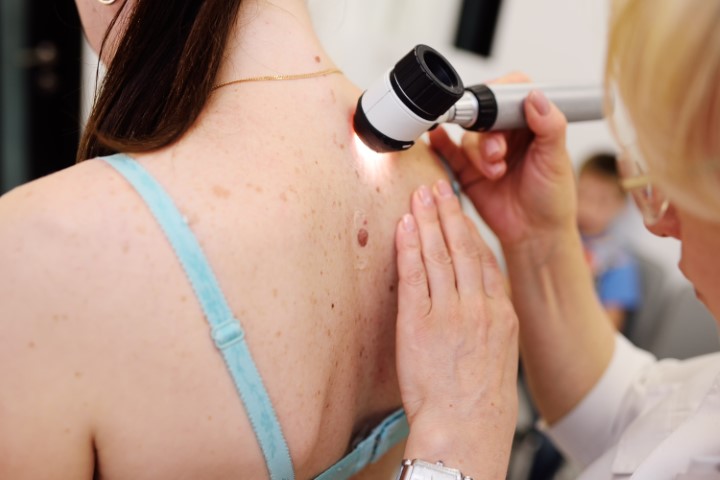
Pigmented skin moles are clusters of melanocytic cells found in the skin. They are benign lesions commonly referred to as moles. They often develop under the influence of sunlight and ultraviolet radiation. An average person has approximately 10 to 40 moles on their skin. The vast majority do not require medical treatment. However, some moles may change over time and develop features that require specialist assessment.
A relatively simple method used to identify suspicious skin lesions and possible melanoma is the ABCDE rule:

If any of these changes occur, regardless of how long the symptoms have been present or how small the lesion is, medical assessment is recommended.
Melanoma is a malignant skin cancer arising from pigment-producing cells called melanocytes. Exposure to ultraviolet (UV) radiation is one of the major risk factors for melanoma. The risk increases with cumulative exposure to sunlight and artificial UV radiation from tanning beds over a lifetime.


People at increased risk of developing melanoma include those with:
People with these risk factors should take particular care to avoid excessive sun exposure and should avoid the use of tanning beds.
Skin cancers are among the most common malignant tumors, while melanoma accounts for only a proportion of all skin cancers.

The basic examination for suspected melanoma is a careful inspection of the skin by a medical specialist. Dermoscopy is a valuable diagnostic tool that allows the doctor to assess the features of a mole and estimate the risk that a skin lesion may represent melanoma.
At our Medical Centre, dermoscopic assessment is performed by Dr Kamil Drucis, a specialist in surgical oncology. Dermoscopy, like any imaging assessment, helps estimate risk, but the definitive diagnosis of a suspicious skin lesion is based on microscopic examination by a specialist pathologist.
Histopathological examination of the completely removed lesion provides the definitive diagnosis and determines the nature of the mole or skin lesion.
When a skin lesion is suspicious for melanoma, the preferred diagnostic approach is complete surgical removal of the lesion for histopathological examination. Suspicious pigmented lesions should not be destroyed by methods that prevent subsequent pathological examination, such as laser treatment or cryotherapy.
A suspicious mole should be removed completely with an appropriate margin of surrounding tissue by performing an excisional biopsy. Complete excision allows the entire lesion to be assessed microscopically and provides the information required for further treatment planning.
Early-stage melanoma is often highly treatable with surgery. Once the disease becomes locally advanced or spreads to lymph nodes or distant organs, treatment becomes more complex and may require a combination of surgical and systemic therapies. This is why early assessment and removal of suspicious skin lesions are so important.

At Tartaczna 2 Medical Centre, excisional biopsies of suspicious skin lesions are performed by surgical oncologists. Every excised suspicious lesion is submitted for histopathological examination.
We invite you to book an appointment with Dr Dawid Śliwiński, Dr Natalia Spychalska or Dr Kamil Drucis through our website (e-mail or telephone contact) or via ZnanyLekarz: Dr Natalia Spychalska, Dr Kamil Drucis.
Please note that the waiting time for a histopathology result is approximately 30 days.