Tartaczna 2


 back
back
Thyroid Nodules in Gdańsk – Assessment and Treatment
Thyroid ultrasound, assessment for biopsy and interpretation of results
Thyroid nodules are focal changes within the thyroid gland. They may be solid, fluid-filled or a mixture of both. Most are benign, and many are found incidentally during an ultrasound scan. Finding a nodule does not mean that you have cancer or need an operation.
At Tartaczna 2 Medical Centre in Gdańsk, thyroid nodule assessment includes an endocrinology consultation, ultrasound and, when indicated, a fine-needle aspiration biopsy. Together, these findings help determine whether monitoring or further treatment is needed.
Thyroid nodule symptoms – when should you see a doctor?
Small nodules usually cause no symptoms. Larger nodules may be felt in the neck, cause a sensation of pressure or make swallowing difficult. Some nodules produce excess thyroid hormones and may cause symptoms of an overactive thyroid, such as palpitations, excessive sweating or weight loss.
A rapidly growing nodule, persistent unexplained hoarseness or enlarged lymph nodes in the neck require prompt assessment. Increasing difficulty breathing requires urgent medical attention.
Thyroid nodule assessment – where to start
The doctor asks about your symptoms, previous investigations, family history of thyroid disease and risk factors, such as previous radiation treatment to the neck. Initial assessment includes a clinical examination, ultrasound of the thyroid and neck lymph nodes, and a TSH blood test.
Depending on the findings, additional blood tests, such as free T4 and free T3, may be needed. If TSH is low, the doctor may recommend thyroid scintigraphy, a nuclear medicine scan that helps determine whether a nodule is producing hormones independently. A normal TSH result does not rule out a thyroid nodule or cancer within it.

Thyroid ultrasound – what does EU-TIRADS-PL mean?
Ultrasound assesses a nodule's size, composition, echogenicity, margins and any suspicious features. The 2022 Polish recommendations advise using EU-TIRADS-PL, the Polish adaptation of the European risk assessment system. The ultrasound category helps determine whether a biopsy is indicated, but it is not a diagnosis of cancer.
| Category | Meaning of the ultrasound findings | Usual biopsy indications under EU-TIRADS-PL |
|---|---|---|
| 1 | No nodules. | Not applicable. |
| 2 | Benign appearance: a pure cyst or an entirely spongiform nodule. | A diagnostic biopsy is usually unnecessary. A cyst causing symptoms may need drainage. |
| 3 | Low risk: an isoechoic or hyperechoic nodule with a regular shape and smooth margins, without high-risk features. | Biopsy at a size of at least 20 mm. |
| 4 | Intermediate risk: a mildly hypoechoic nodule with a regular shape and smooth margins, without high-risk features. | Biopsy at a size of at least 15 mm. |
| 5 | High risk: at least one highly suspicious feature, such as irregular margins, microcalcifications, marked hypoechogenicity or suspected extension beyond the thyroid. | Biopsy at a size of at least 5 mm. |
This table refers specifically to EU-TIRADS-PL, rather than every TIRADS system. The decision also takes account of lymph node findings, test results and individual risk factors. In certain circumstances, a biopsy may be needed regardless of nodule size. If an ultrasound report does not include a category, this does not automatically mean a biopsy is required: the doctor reviews the full report and arranges further assessment if necessary.

When is a thyroid fine-needle aspiration biopsy performed?
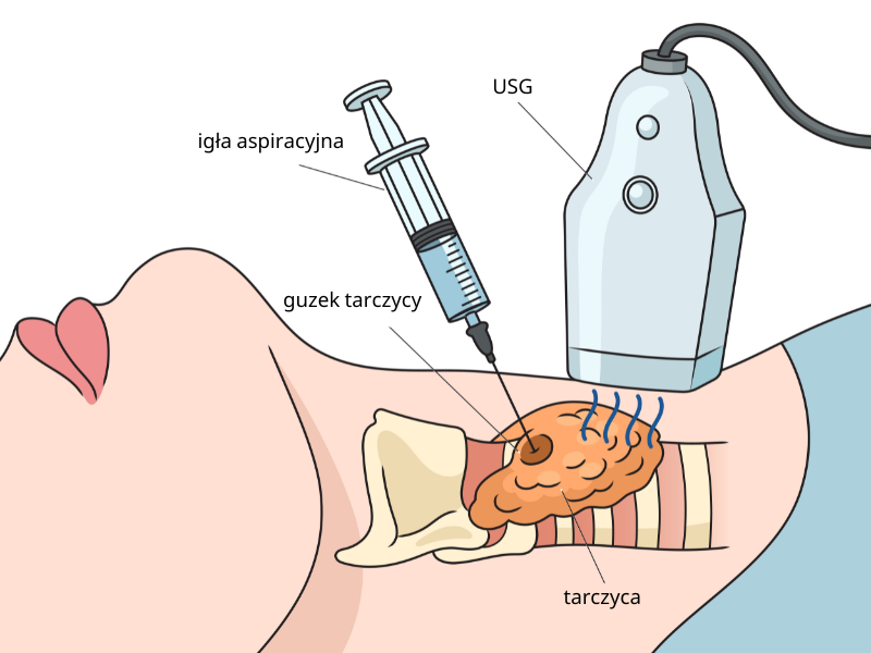
Ultrasound-guided fine-needle aspiration, or FNA, involves taking cells from a selected nodule. A pathologist examines the sample using cytology. Biopsy indications depend on the nodule's features and size, together with the clinical circumstances. In Polish medical reports, the procedure may be abbreviated as BAC or BACC.
When there are several nodules, those requiring further assessment are selected for biopsy, with particular attention to suspicious features. The result from one nodule does not establish the nature of all the others. For details of preparation and the procedure, see our page on thyroid fine-needle aspiration biopsy in Gdańsk.
Thyroid biopsy results – Bethesda categories
The Bethesda system is used to report thyroid cytology results. The 2023 edition defines six categories. Results should be interpreted alongside the ultrasound findings and clinical information.
| Category | Meaning and further management |
|---|---|
| I – nondiagnostic | The sample does not allow a reliable assessment. A repeat ultrasound-guided biopsy is usually needed; the next steps depend on the nature of the nodule. |
| II – benign | Monitoring is usually appropriate. Suspicious ultrasound findings or significant changes on subsequent examinations may justify another biopsy. |
| III – atypia of undetermined significance | Some features prevent the nodule from being classified with confidence. A repeat biopsy is usually recommended; molecular testing or surgery may be considered in selected cases. |
| IV – follicular neoplasm | Cytology cannot determine whether the lesion is benign or malignant. A specialist consultation is needed to discuss further assessment, often involving surgery. |
| V – suspicious for malignancy | Prompt specialist assessment is required to agree a treatment plan, which most often involves surgery. |
| VI – malignant | The cytological diagnosis indicates malignancy. Further management depends on the type of tumour, its extent and the patient's health. |
The categories describe different levels of risk and are not cancer stages. A nondiagnostic result does not mean that a nodule is benign, and category III or IV is not equivalent to a cancer diagnosis.
What does Bethesda IV, V or VI mean – is surgery necessary?
One limitation in category IV is that cells alone cannot reliably show whether a tumour has invaded its capsule or blood vessels. Histopathological examination of tissue removed during surgery may therefore be essential for distinguishing a follicular adenoma from follicular carcinoma. Molecular testing can be helpful in selected circumstances; the doctor will discuss its usefulness and availability.
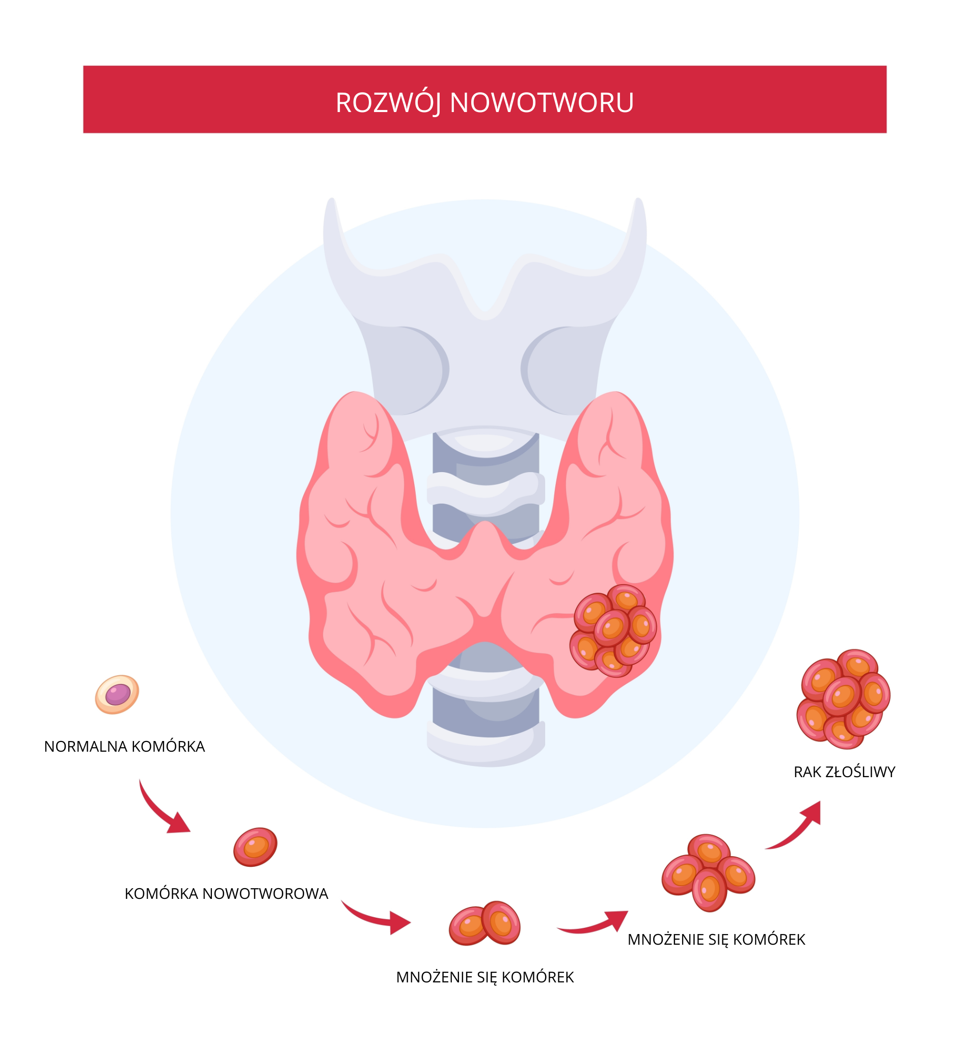
Category V and VI results most often lead to assessment for surgery, but management depends on the type of tumour. For selected patients with low-risk papillary thyroid microcarcinoma, an experienced team may offer active surveillance. This involves scheduled follow-up according to a defined protocol. The decision cannot be made solely from the Bethesda category number.
Monitoring and treatment of benign thyroid nodules
Benign nodules that do not cause symptoms or hormonal disturbances often need monitoring alone. The timing of the next ultrasound depends on the risk category, previous cytology, nodule size and changes over time. Not every nodule needs an annual biopsy.
Nodule growth does not necessarily mean cancer, but it may warrant reassessment. Treatment may be considered if a nodule compresses nearby structures, causes swallowing difficulties or an overactive thyroid, or shows a significant change in its ultrasound appearance.
Thyroid cancer – what affects the outlook?
The outlook depends on the type and stage of the tumour, the patient's age and the response to treatment. Many differentiated thyroid cancers, particularly papillary cancer detected at an early stage, have a very favourable prognosis. This does not apply equally to every type of thyroid cancer, so each case requires an individual assessment.
Thyroid nodules in Gdańsk – doctors and appointments
At Tartaczna 2 Medical Centre, you can book:
- thyroid ultrasound with Dr Ewa Kołyszko – assessment of focal thyroid changes and neck lymph nodes,
- an endocrinology consultation with Dr Anna Florysiak – assessment and planning of further management,
- ultrasound-guided thyroid biopsy with Dr Kamil Drucis – once biopsy indications have been established,
- an oncology consultation with Dr Magdalena Korożan – when the findings indicate that this is needed.
Bring previous ultrasound reports and images, biopsy results, blood test results and a list of your medicines. These help the doctor compare changes over time and plan the next steps.
Doctor profile: Dr Kamil Drucis.
Book using the appointment calendar below or contact our reception team: +48 58 719 10 25, kontakt@tartaczna2.pl.