
Tartaczna 2


 back
back
Breast Lump on Ultrasound – BI-RADS Classification
Breast Lump and the BI-RADS Scale
Breast cancer is the most common cancer in adult women. The peak incidence occurs between the ages of 55 and 65. However, breast cancer can also occur in younger women. It may be associated with a family history of cancer. Hereditary breast cancer can be identified through genetic testing. Genetic profiling for cancer predisposition can be performed using blood or saliva samples, while molecular testing may also be carried out on tumour tissue.
Mammography and Breast Ultrasound – Assessment of Breast Lumps and Tumours
The main imaging methods used to assess breast lumps, tumours and other breast lesions include mammography, breast ultrasound and, in selected cases, magnetic resonance imaging (MRI). The radiological characteristics of a breast lesion are assessed by a radiologist using the BI-RADS scale. BI-RADS, or the Breast Imaging Reporting and Data System, is a standardised system for reporting breast imaging findings. Every breast imaging report should include a BI-RADS assessment of the identified lesions. At our Medical Centre, we perform comprehensive breast ultrasound examinations using a new-generation LOGIQ E10 ultrasound system equipped with elastography and Power Doppler (PD). Breast ultrasound examinations are performed by radiologists and may also be performed by a surgical oncologist during a specialist consultation. Women from the general population should undergo regular breast examinations once a year.
In menstruating women with predominantly glandular breast tissue, breast ultrasound is recommended. Mammography before the age of 50 has more limited diagnostic value, particularly in women with dense or glandular breasts. In women over the age of 50, mammography is routinely performed every 2 years, together with breast ultrasound examinations. Since 2023, women aged 45–74 have been eligible for screening mammography without a referral as part of the national breast cancer screening programme.
Have you received an abnormal BI-RADS result or has a breast lump or tumour been found on imaging? Further diagnostic assessment can be carried out at our Breast Clinic in Gdańsk, where, depending on your imaging results, specialist consultations, breast ultrasound and appropriately selected breast biopsy procedures are available.
BI-RADS Scale
- BI-RADS 0 - the examination is inconclusive and requires additional breast imaging using another diagnostic method.
- BI-RADS 1 - normal result, with no abnormal findings. Routine breast examination once a year is recommended.
- BI-RADS 2 - benign findings with no features suggesting malignancy, e.g. fibrocystic changes, cysts or stable breast lesions that do not change during follow-up. Breast examination (mammography or ultrasound) once a year is recommended.
- BI-RADS 3 - a probably benign breast lesion has been detected. The risk of malignancy is up to 2%. This category may include findings such as clusters of microcalcifications, complicated cysts, fibroadenomas or asymmetric densities. Early imaging follow-up is recommended, typically breast ultrasound after 6 months. In women at increased risk of breast cancer, fine-needle aspiration biopsy (FNA) or core needle biopsy (CNB) may be recommended to obtain material for cytological or histopathological examination.
- BI-RADS 4 - a suspicious breast lesion or tumour requiring biopsy has been identified in order to determine its nature. This category indicates suspicion of malignancy. Depending on the estimated risk of malignancy, BI-RADS 4 is divided into the following subcategories:
- BI-RADS 4a - risk of malignancy 2–10%
- BI-RADS 4b - risk of malignancy 10–50%
- BI-RADS 4c - estimated risk of malignancy 50–95%.
In every case of BI-RADS 4, histopathological verification of the breast lesion by core needle biopsy or vacuum-assisted breast biopsy is required.
- BI-RADS 5 - an imaging examination showing a breast lesion highly suspicious for malignancy, with a risk of cancer greater than 95%. Urgent core needle biopsy is recommended. A negative histopathological result does not completely exclude malignancy and does not eliminate the need for additional diagnostic assessment and/or surgical biopsy.
- BI-RADS 6 - indicates breast cancer that has already been diagnosed and confirmed by histopathological examination.
When a breast tumour, breast lump or other breast lesion is identified on imaging, its nature generally needs to be established before treatment is started. The most common breast lesions are benign findings such as fibroadenomas and breast cysts. However, some benign and malignant breast tumours and lesions may appear similar, particularly when they are small, detected incidentally on imaging or occur in younger women. Determining the nature of a breast lump or tumour is particularly important because the earlier breast cancer is detected, the greater the chance of successful treatment.

Comprehensive Diagnosis of Breast Tumours at Tartaczna 2 Medical Centre in Gdańsk
- Breast ultrasound examinations and imaging assessment of breast lumps and other breast lesions are performed by radiologists Dr Ewa Kołyszko and Dr Jakub Morawiec.
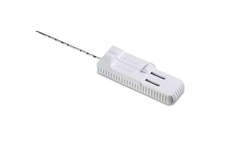
- In patients with a previously identified breast lump or tumour, further diagnostic assessment may include FNA (fine-needle aspiration biopsy), CNB (core needle biopsy), vacuum-assisted breast biopsy and surgical excision of the breast tumour. These procedures are performed by surgical oncologists Dr Natalia Spychalska and Dr Kamil Drucis. Radiologist Dr Jakub Morawiec also performs breast biopsies.
- Patients diagnosed with a breast tumour, including breast cancer, who require consultation regarding treatment options, further diagnostics and oncological management are invited to consult clinical oncologist and radiation oncologist Dr Magdalena Korożan.
Book an appointment at our Breast Clinic in Gdańsk through our website (by e-mail, telephone or online registration) — our doctors: Dr Ewa Kołyszko, Dr Jakub Morawiec, Dr Kamil Drucis, Dr Natalia Spychalska, Dr Magdalena Korożan, Dr Artur Antolak.
Please note that the waiting time for a histopathology report is approximately 30 days.

Breast Fine Needle Aspiration Biopsy (FNA)
Breast Core Needle Biopsy
