Tartaczna 2


 back
back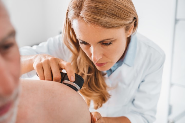
Dermoscopy in Gdańsk – Mole and Skin Lesion Checks
Assessment of moles and suspicious skin lesions
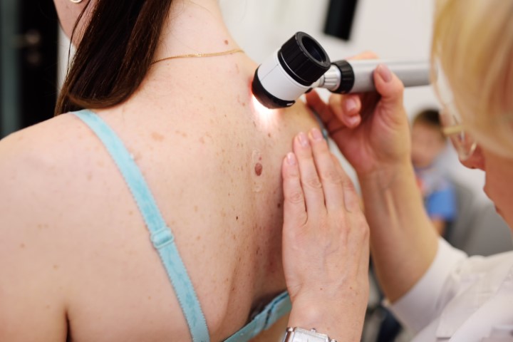
Dermoscopy, also known as dermatoscopy, is a non-invasive examination of moles and other skin lesions. A doctor uses a dermoscope to view the skin under magnification and assess details that are not visible to the naked eye. The examination helps identify lesions that need further investigation, including those suspicious for melanoma.
At Tartaczna 2 Medical Centre in Gdańsk, we examine moles and other skin lesions and assess whether they should be monitored or removed for histopathological examination.

When should you have your moles checked?
A consultation is particularly advisable if:
- you have a new skin lesion that is growing or looks different from the others;
- an existing mole changes in shape, colour, size or surface appearance;
- a lesion bleeds without an obvious injury, itches, becomes ulcerated or does not heal;
- you have numerous moles or atypical moles;
- you or a close relative have been diagnosed with melanoma;
- you are planning to have a mole removed, including for cosmetic reasons.
A change in a mole's appearance does not necessarily mean cancer, but it should be assessed. If a lesion is growing rapidly or bleeding, do not wait until your next routine check-up.
What happens during a dermoscopy examination?
The appointment begins with a discussion of your skin lesions, how they have changed, any previous procedures and any family history of skin cancer. The doctor examines your skin and assesses moles using a dermoscope. Comparing a lesion with your other moles and examining the skin across your whole body are important parts of the assessment.
A dermoscope uses magnification and specialised lighting. Depending on the device, the examination may involve contact with the skin and the use of a contact fluid, or it may use polarised light. The doctor assesses features including symmetry, pigment distribution, structures within the lesion and blood vessels.
The examination does not involve cutting the skin and does not require anaesthesia. The length of the appointment depends on the number of lesions and the scope of the assessment. Afterwards, the doctor explains the findings and recommendations.
How should you prepare for dermoscopy?
- You do not need to fast.
- Do not cover the lesions being examined with make-up or tinted cosmetic products.
- If your nails need to be examined, remove nail polish or artificial nail coverings so that the nail plate is visible.
- Bring any previous dermoscopy reports, histopathology results and photographs showing changes over time.
- Point out any moles that have changed in appearance or are causing symptoms.
Can dermoscopy detect melanoma?
Dermoscopy improves the accuracy of the clinical assessment of skin lesions and helps identify those suspicious for melanoma. However, it cannot confirm or exclude cancer with complete certainty. A definitive diagnosis of a suspicious lesion is based on histopathological examination of a tissue sample.
The examination is also useful for assessing other lesions, including other skin cancers and benign changes. Not every melanoma is dark in colour, and not every melanoma develops from an existing mole.
Understanding dermoscopy results and the TDS score
Doctors may use different methods to analyse dermoscopic images. One is the ABCD rule of dermoscopy, which is used to calculate the total dermoscopy score, or TDS. It considers the lesion's asymmetry, borders, colours and visible structures.
The TDS has limitations and is not suitable for every type of lesion or every body site. A low score does not rule out melanoma, and the decision to remove or monitor a lesion must take the full clinical picture into account. The score alone should not determine whether further investigation is deferred.
Melanoma warning signs – the ABCDE rule
The ABCDE rule can help you notice features that should be checked by a doctor. It is not a test for diagnosing melanoma yourself.
| Feature | What should you look for? |
|---|---|
| A – Asymmetry | One part of the lesion differs from the other in shape or appearance. |
| B – Border | The edges are irregular, notched or poorly defined. |
| C – Colour | The lesion has uneven pigmentation or several different colours. |
| D – Diameter | A diameter greater than 6 mm deserves attention, but melanoma can also be smaller. |
| E – Evolving | The mole is changing in size, shape, colour or appearance. |
Even one concerning feature is a reason to seek a consultation. A mole that looks noticeably different from your others, known as the “ugly duckling” sign, also deserves attention. Melanoma may not show all the ABCDE features.
How often should you have your moles checked?
Your doctor recommends the frequency of examinations based on the number and type of moles, previous findings and your individual risk. People with numerous atypical moles, a personal history of melanoma or a family history of melanoma may need more frequent checks.
If a selected atypical lesion does not require removal at the first assessment, the doctor may recommend a short-term review, for example after around 3 months, with comparison of photographic records. A lesion suspicious for melanoma requires prompt investigation rather than routinely waiting several months.
Between appointments, check your skin, including your back, scalp, palms, soles and nails. New concerning symptoms are a reason to seek an earlier assessment, regardless of when your next routine check is scheduled.
When does a mole need to be removed?
If a lesion is suspicious for melanoma, the usual recommendation is to remove it completely with a small margin and send it for histopathological examination. The doctor decides how the tissue should be sampled. You can find more information on our page about surgical removal of suspicious skin lesions.
Lesions suspicious for melanoma should not be destroyed using laser treatment, freezing or electrocoagulation, as this prevents proper histopathological assessment of the entire lesion. Moles considered benign do not always need to be removed.
Dermoscopy in Gdańsk – book a mole check
At Tartaczna 2 Medical Centre, dermoscopy examinations are performed by:
- Sara Strugała, MD;
- Kamil Drucis, MD, PhD – surgical oncologist.
You can find information about fees in our dermatology price list. Reception can also confirm the price and scope of your chosen appointment.
Book through Tartaczna 2 Medical Centre reception, call +48 58 719 10 25 or email kontakt@tartaczna2.pl.