Tartaczna 2


 back
back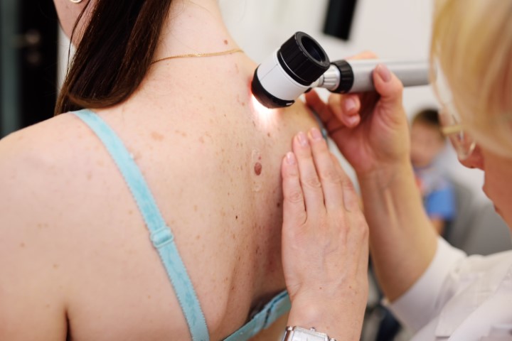
Mole Removal in Gdańsk – Melanoma Assessment
Mole checks and surgical removal of suspicious skin lesions
Mole removal in Gdańsk at Tartaczna 2 Medical Centre begins with an assessment of the lesion to determine whether it needs monitoring or excision for histopathological examination. Consultations for suspicious moles and other skin lesions are provided by Dr Kamil Drucis, Dr Natalia Spychalska and Dr Sara Strugała.
What are pigmented naevi, commonly known as moles?
Pigmented naevi are usually benign skin lesions formed by clusters of melanocytes – the cells that produce skin pigment. They may be present at birth or develop later in life. Genetic factors and exposure to ultraviolet (UV) radiation both contribute to their development.
Most moles do not need to be removed. However, it is worth checking your skin for new, unusual lesions or changes in existing moles. Melanoma can also develop in an area where there was no mole before.
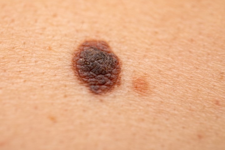
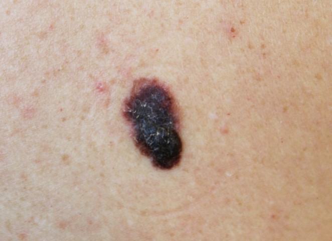
When should a mole cause concern? The ABCDE rule
The ABCDE rule helps identify features that need medical assessment:
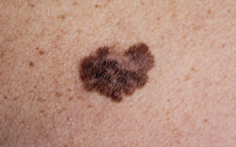
- A – asymmetry: one part of the lesion differs in shape or appearance from the other.
- B – border: the edges are uneven, notched or poorly defined.
- C – colour: the lesion has uneven colouring, such as different shades of brown, black, red, white or blue.
- D – diameter: a lesion larger than approximately 6 mm deserves particular attention, but melanoma can also be smaller.
- E – evolution: the lesion is growing or changing in shape, colour, surface appearance or other features.

A lesion that looks noticeably different from your other moles, a rapidly growing nodule, spontaneous bleeding or a non-healing ulcer also needs assessment. A lesion does not have to meet all the ABCDE criteria for you to see a doctor. A small size and the absence of pain do not rule out a serious condition.
Mole checks and dermoscopy
The doctor assesses the appearance of the lesion, how long it has been present and any changes such as growth, bleeding or other symptoms. During dermoscopy, the skin is examined under magnification with specialised lighting, allowing structures that are invisible to the naked eye to be seen.
The examination helps determine whether a mole can be monitored or needs to be removed. Dermoscopy does not replace histopathological examination when a lesion is suspicious for cancer. Follow-up intervals are agreed individually; a new or changing lesion needs earlier assessment.
When is surgical mole removal needed?
Surgical mole removal is considered primarily when the appearance or evolution of a lesion raises concern about cancer. The doctor decides whether removal is indicated after an examination, taking into account the location of the lesion and your medical history.
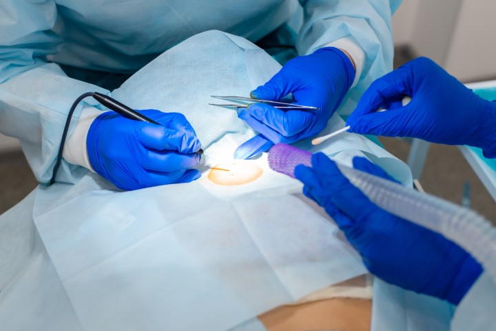
When melanoma is suspected, the preferred approach is to remove the entire lesion with a narrow margin of surrounding skin and send it for histopathological examination. This is called an excisional biopsy. We describe preparation, the procedure and healing on a separate page.
In selected situations, such as a large lesion or a difficult location, the doctor may plan to take a sample of part of the lesion. An appropriately chosen biopsy is part of melanoma diagnosis and should not be delayed out of concern that taking a sample will itself cause the cancer to spread.
Removing all moles as a precaution is not recommended. Having one lesion removed does not replace monitoring the rest of your skin.
Can a suspicious mole be removed with a laser?
A lesion suspected of being melanoma should not be destroyed with a laser, frozen or cauterised. These methods may prevent proper histopathological assessment and delay diagnosis. A medical assessment is needed before choosing how to remove a mole.
For benign skin lesions, treatment is selected according to the type of lesion. Methods suitable for certain benign growths may not be appropriate for pigmented moles.
What is melanoma, and who is at greater risk?
Melanoma is a malignant tumour that develops from melanocytes. It can arise within an existing mole or on skin where no lesion was previously visible. Not every melanoma is dark or looks like a typical mole.
Factors that increase the risk include:
- exposure to UV radiation, sunbed use and previous sunburns,
- fair skin that burns easily,
- numerous or atypical moles,
- a personal or family history of melanoma,
- a weakened immune system, including immunosuppression caused by treatment.
Protecting your skin includes avoiding sunburn and sunbeds, seeking shade, wearing suitable clothing and using sunscreen. People at higher risk should agree an individual mole-check schedule with their doctor.
Histopathological examination of a removed mole
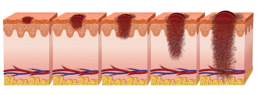
Every suspicious lesion removed for diagnosis at our centre is sent for histopathological examination. A pathologist determines the nature of the lesion and, if melanoma is diagnosed, reports the features needed to plan further treatment, including Breslow thickness, ulceration and excision margins.
The waiting time for histopathology results is approximately 30 days. Additional staining or a specialist review of the tissue slides may extend this timeframe. Discuss your results with the doctor managing your assessment.
What happens if the results confirm melanoma?
A melanoma diagnosis requires a plan for further management. Following a diagnostic biopsy, further surgery to remove additional tissue around the scar is usually needed. A sentinel lymph node biopsy may also be considered for some patients. The extent of treatment depends mainly on the histopathology findings and the stage of the disease.
Early diagnosis improves the chances of successful treatment. Depending on the circumstances, treatment may include surgery, immunotherapy or molecularly targeted therapy. Prognosis cannot be determined from the appearance of a mole alone.
If an oncology consultation is indicated to discuss the results and further treatment, you can book an appointment with Dr Magdalena Korożan at our centre.
Mole removal in Gdańsk – doctors and appointments
For mole assessment and advice on treatment at Tartaczna 2 Medical Centre, book an appointment with:
- Dr Kamil Drucis – surgical oncology,
- Dr Natalia Spychalska – surgical oncology,
- Dr Sara Strugała – dermatology.
Excisional biopsies of lesions suspected of being melanoma are performed by surgical oncologists at our centre. Bring any previous dermoscopy or histopathology reports to your consultation, if available.
Book using the appointment calendar below or contact our reception team: +48 58 719 10 25, kontakt@tartaczna2.pl. When booking, please specify whether you need a mole check or a consultation before lesion removal.